Why Choose Baptist Health?
At the UAB Medicine Breast Health Clinic, part of the UAB Medicine Multispecialty Clinic at Baptist Medical Center South, our compassionate team is dedicated to providing comprehensive, personalized breast care to the women of Central Alabama. With empathy and encouragement guiding our approach, we specialize in meeting the unique needs of women and their families. We offer clear explanations every step of the way through the breast surgery process.
Our breast surgeons collaborate closely with your other providers to develop a customized treatment plan tailored to your individual needs. Through continual follow-up and unwavering support when you need it most, we strive to provide the highest level of state-of-the-art, convenient care close to home with your loved ones by your side.
While new patient referrals are required, our team is glad to guide you through the process and address any questions you may have. We are committed to delivering prompt, comprehensive, compassionate care in a comfortable, supportive environment.
Breast health issues can be challenging. Our team is here with a guiding hand to help.

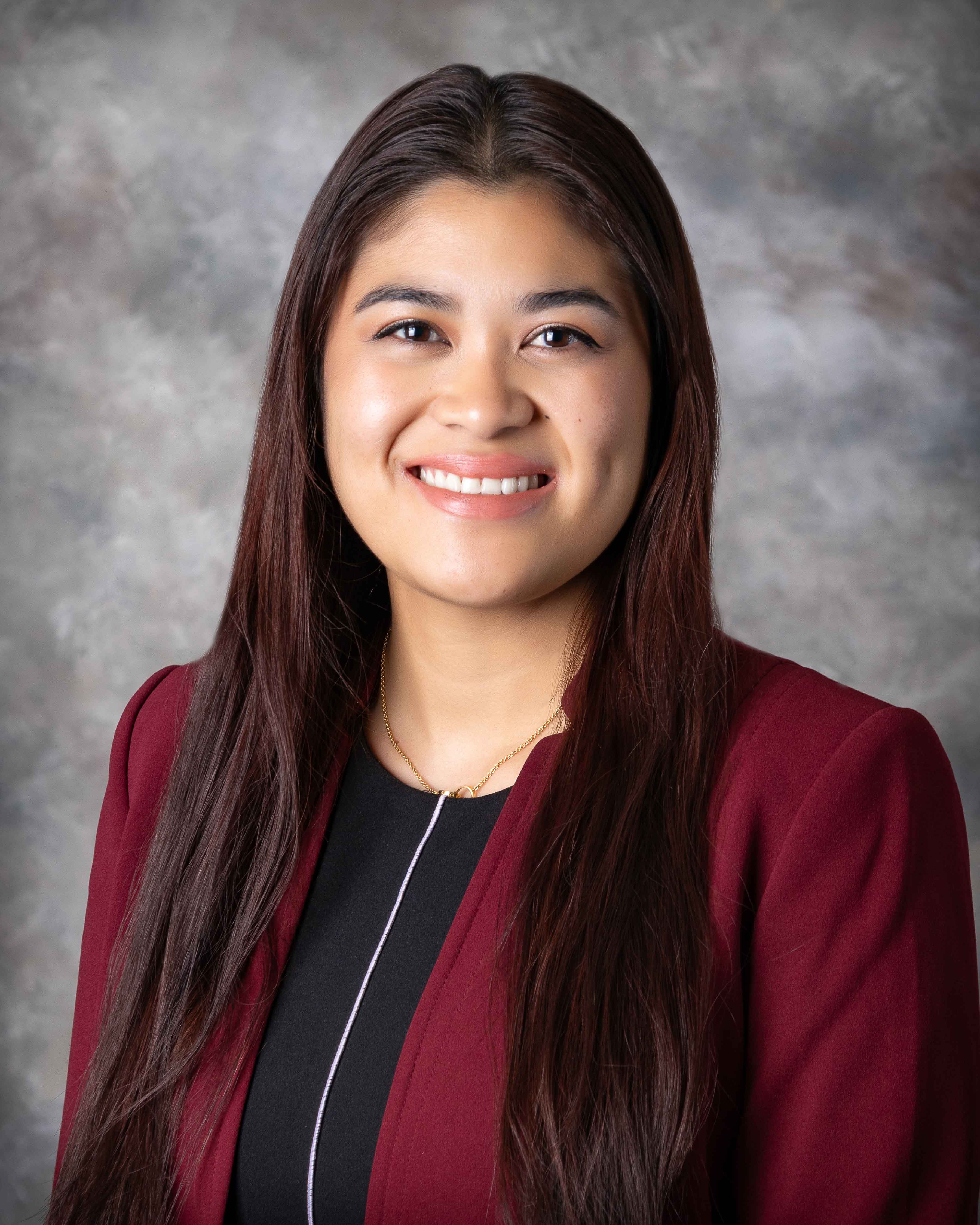
Sasa Espino, MD, FACS
Dr. Sasa Espino received her medical degree from the Virginia Commonwealth University (VCU) School of Medicine in Richmond, Virginia. She completed her General Surgery residency at VCU School of Medicine. She also completed a Breast Surgery fellowship at the Northwestern University Feinberg School of Medicine in Chicago, Illinois.
Board certified by the American Board of Surgery, Dr. Espino’s clinical interests and procedures include cancerous and non-cancerous Breast Surgery.
A proud member of the American Society of Breast Surgery, American College of Surgeons, Society of Surgical Oncology and American Medical Association, she is certified in Oncoplastic Breast Surgery.
Dr. Espino enjoys mountain biking, snowboarding, surfing, learning new languages and playing the piano in her spare time.

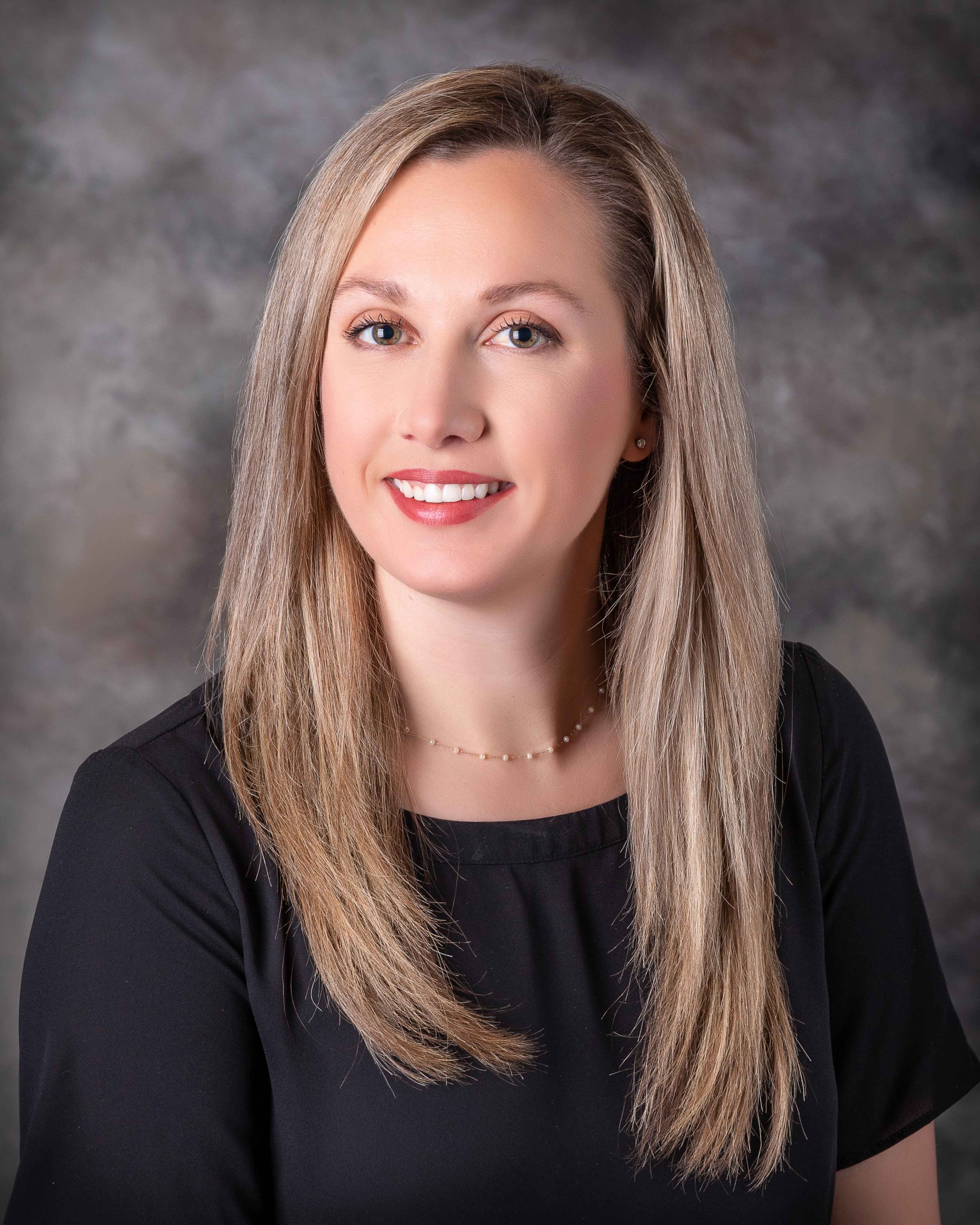
Roni Alcantara, PA-C
Ms. Alcantara received her master’s degree in physician assistant studies from the University of Alabama at Birmingham in Birmingham, Alabama. She also has a bachelor’s and master’s degree in public health from the University of Alabama at Birmingham.
As a certified Physician Assistant, Roni has clinical interests in Breast Surgery, Oncology, Counseling, and Public Health. In her spare time, she enjoys traveling, playing volleyball, and dining out at her favorite restaurants.
She looks forward to serving the people of the River Region and surrounding areas, providing them with patient education, pre-operative and post-operative care, and assessing and managing patients with various breast conditions.

Kristine Bauer, PA-C
Ms. Bauer received her Master's in Physician Assistant Studies from Faulkner University in Montgomery, Alabama and a Bachelor of Science Degree in Biology from the University of Louisiana in Lafayette, Louisiana.
A proud member of the American Academy of Physician Associates (AAPA), Kristine has clinical interests in assisting in surgical procedures, Breast Cancer Risk Assessment and Counseling, as well as Genetic testing for the BRCA gene and other mutations associated with Breast Cancer.
She focuses on continuous follow-ups and surveillance for both cancer and high-risk patients.
Board certified with the National Commission on Certification of Physician Assistants, Kristine was awarded the Surgical Excellence Award, which recognizes high-quality work in surgical service. In her spare time, she enjoys spending time with her husband and dog, talking to her mother, hiking and cooking.
Conditions and Treatments
An abscess is a localized area of infection with a walled-off collection of pus. An abscess is generally treated with antibiotics and drainage. Drainage may be performed with a needle and syringe (aspiration).
Approximately 1 in 8 women will develop breast cancer in their lifetime. There are multiple different risk models that can help estimate a woman’s individual risk of breast cancer, taking in consideration factors such as family history, use of hormones, breast density, and history of previous breast biopsies. None of the risk models can tell whether or not a person will develop breast cancer.
Breast ultrasound uses high-frequency sound waves to produce pictures of the internal structure of your breast tissue. It does not involve radiation. Breast ultrasound is primarily used to assess abnormalities detected during physical exam or to characterize abnormalities seen on a mammogram. Breast ultrasound cannot take the place of mammography for breast cancer screening. A screening study is one that is performed on a large number of people in order to identify those who have or are likely to develop a disease or condition.
Family history is a very important risk factor for breast cancer. However, only 5-10% of all breast cancers are thought to be hereditary, or caused by abnormal genes passed down from one generation to another. Women who meet the following criteria may consider genetic testing:
- First- or second-degree relative diagnosed with breast cancer at an early (age 45 or under)
- Ashkenazi Jewish heritage and family history of breast cancer
- Two or more breast cancers in a single family member
- Family or personal history of ovarian cancer, fallopian cancer, or primary peritoneal cancer
- Male breast cancer
- Known genetic mutation carrier in the family.
Genetic testing in our office is done through a blood or spit test. The test extracts DNA from the blood or the cells that are shed in your saliva. The DNA is analyzed to look for mutations (or changes) in the DNA that may be related to cancer risk.
Women who are at increased risk for breast cancer may benefit from high risk follow up to provide structured surveillance and education. The goal of high risk screening is prevention and/or early detection of breast cancer. This may be accomplished through preventive measures, such as medications and lifestyle modifications, and imaging studies, including mammography, tomosynthesis (3D mammography), ultrasound, and breast MRI.
A cancer diagnosis can be confusing and frightening for both the patient and family members/caregivers. It is important to find a caring physician with expertise in your type of cancer. Asking for a second opinion is a fairly common practice and may provide the following:
- Confirmation of a diagnosis
- Information about other treatment options
- A sense of comfort and confidence with your healthcare decisions
This procedure uses a syringe to withdraw fluid from a cyst. A cyst is a fluid- filled sac within the breast tissue that may look or feel like a lump. This procedure is reserved for cysts that are large or painful.
This is a type of breast biopsy that uses a needle to remove a small sample of tissue from a solid mass. The tissue is then sent to a lab for analysis to determine a diagnosis. A local anesthetic is used to numb the skin and breast tissue at the biopsy site. A small nick is made in the skin, and ultrasound is used to guide the biopsy needle into the mass. Several samples of tissue, each about the size of a grain of rice, are removed. A tiny titanium marker may be inserted to identify the biopsy site so it can be located in the future if necessary. The procedure is usually completed within an hour. You do not need a driver but should avoid strenuous activity for 24 hours after the procedure. You will be given an ice pack for comfort and may take an over-the- counter pain medication such as Tylenol or Advil. Mild swelling and bruising are normal after the procedure.
Insertion of marking clip: A tiny titanium clip is inserted to mark the biopsy site. If the biopsy is benign, the clip will remain in the breast but should not cause any trouble. It will not set off scanners in the airport, and you can still have an MRI with the clip in place. If the mass turns out to be a cancer, the clip marks the spot that will eventually need to be removed. The clip can usually be seen with ultrasound, allowing us to make an incision right over the cancer in the operating room.
At times, an abnormality may appear on mammogram that cannot be felt or seen on ultrasound. Traditionally, wire localization was the preferred preoperative technique for localizing breast tumors; however, we utilize SAVI SCOUT® utilizing a reflector that's about the size of a grain of rice in lieu of a wire. With placement inside the tumor up to 30 days before surgery, the reflector remains passive until activated, isn't externally visible, and patient movement is not restricted by it. The nonradioactive surgical guidance technology is activated by your surgeon, guiding them to the precise site of your tumor and increasing the likelihood of total tumor removal.
Since the early 1990s, the preferred surgical treatment for most breast cancer has been breast preservation, frequently referred to as a partial mastectomy or lumpectomy. Many excellent studies in which women were randomly assigned to either mastectomy or partial mastectomy have demonstrated that there is no difference in survival whether a woman chooses mastectomy or breast preservation. The studies were done in the early 1970's, so the follow-up of those patients is very long, and our confidence in the findings is quite high. A partial mastectomy consists of removing the cancer and a small rim of normal tissue around it to be sure all of the cancer has been removed. We usually use ultrasound in the operating room to help us know what tissue to remove. An x-ray of the specimen is taken immediately after removing it in order to confirm that the targeted tissue was removed and the tumor is not too close to the cut edge of the tissue (margin). We have a special x-ray machine in the operating room to x-ray the specimen.
A mastectomy is still needed about a third of the time, when the cancer is extensive in the breast and it would be difficult to remove it all without severely deforming the breast. Often, it is difficult to be sure where the borders of the cancer begin and end, making it difficult to know just how much of the breast tissue to remove. Other times, when there is extensive malignant calcification on the mammogram, particularly if it extends up under the nipple, a mastectomy is a better option. Sometimes, when a cancer is localized but too large to allow breast preservation pre-operative (also called "neo-adjuvant" or "induction") chemotherapy or hormonal therapy can may shrink the cancer to allow breast conservation. Numbing medicine is injected around a nerve just above the breast, and this helps significantly with pain after surgery. Mastectomy is usually done as an outpatient. If immediate reconstruction is not planned, information about post-mastectomy bras including an insert to take the place of the missing breast (prosthesis) will be provided.
When a mastectomy is necessary, we encourage women to consider immediate reconstruction, and we will make you an appointment to see a plastic surgeon. It is "reconstructive" and not "cosmetic" surgery, and is therefore covered by essentially all insurance companies. Medicaid does have some restrictions on what they will cover. There are many advantages to immediate reconstruction. The psychological advantage is obvious. When mastectomy is performed, any excess fatty tissue under the arm and extending toward the back may be left behind, resulting in a "pouch of extra tissue.” This is not as noticeable when the breast has been reconstructed. Reconstruction may also be performed at a later date (delayed) if a woman is unsure or requires other treatment. There are two types of reconstruction, autologous (using your own tissue from another area such as the lower abdomen) and tissue expander (using a temporary implant that is injected with saline to stretch the tissue enough to make space for a saline or silicone implant). With immediate reconstruction, the plastic surgeon comes in to begin the reconstruction once the breast tissue has been removed. For tissue expander reconstruction, a second, smaller, procedure is required later to exchange the tissue expander for an implant. The plastic surgeon will determine which type of reconstruction is right for you.
One of the common places breast cancer spreads is to the lymph nodes under the arm, called the axillary nodes. In the past these nodes were always removed in patients with invasive breast cancer in an operation called axillary lymph node dissection. There were several problems with that approach. Many patients (in the range of 25-30% or so) developed a permanent swelling of the arm called lymphedema. In addition, there was a great deal of numbness and discomfort in the axilla (underarm) and upper part of the arm. This procedure has been largely replaced by a technique known as sentinel node biopsy, where a tracer is injected into the breast on the day of surgery to allow identification of the first few lymph node that cancer cells would have spread to if they had spread. These nodes are then removed and analyzed. This procedure has markedly reduces the risk of lymphedema.
Over the past two decades, the way we perform a mastectomy has evolved. In performing a "traditional" mastectomy, much of the overlying skin, as well as the nipple areolar complex, is removed. We now know that it is safe to remove much less skin, and even preserve the nipple areolar complex at times. Studies have demonstrated the safety of skin-sparing and nipple-sparing mastectomy in the setting of cancer, but these procedures may not feasible for all women depending on the size and shape of their breasts.